
- Neo Biotech
- Mar 20
- 2 min read

Updated: May 8

Case Summary 🔎
Patient Information
Age/Sex: 51-year-old Male
Systemic Condition: History of Hypertension and under medication
Clinical Findings
Tooth Site: Mandible
Oral examination: #43 with Grade 3 mobility
Diagnosis: Chronic periodontitis
Clinical Summary
The patient presents with a fully edentulous mandibular arch, except for the mandibular right canine (#43), which exhibits Grade 3 mobility. In the maxilla, a six-unit anterior bridge prosthesis is in place, and implant placement has been performed in both right and left posterior region. A full arch prosthetic rehabilitation is planned for the edentulous mandible.
Treatment Plan
Extraction of #43
Placement of 8 implants in the mandible using Neo NaviGuide for guided surgery
Final restoration with Zirconia crowns
Conclusion
In this case, a fully edentulous mandible was rehabilitated using the Neo NaviGuide system for the placement of 8 implants, followed by guide bone regeneration(GBR). Definitive prosthetic rehabilitation was successfully completed 8 months post-surgery. The patient demonstrated a stable outcome with good function and prognosis over an 7 year follow up.
Case Presentation
1️⃣ Pre-Op
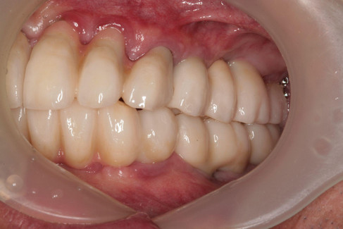
The pre-operative panoramic radiograph and intraoral photograph show a fully edentulous mandibular arch, except for #43.
#43 exhibits severe mobility (Grade 3) and significant bone loss. Generalized alveolar ridge resorption is evident, indicating the treatment for full-arch implant-supported rehabilitation for function and esthetic restoration.


2️⃣ Treatment Plan


Neo NaviGuide fabrication

Neo NaviGuide drilling


3️⃣ Surgery - Full Arch Rehabilitation
The Neo NaviGuide was placed in the mouth. The gingiva at the surgical site is removed using Tissue punch for flapless surgery.
Initial drilling is performed to assess bone density and ensure accurate positioning for implant placement.


Step by step drilling is performed while assessing bone density. Bone particles collected during drilling can be used to autogenous bone.
Based on the bone density, cortical tapping is performed in dense cortical bone to achieve maximum BIC and optimal fixation.


Bone graft is performed using PRF, Regenoss (Allograft) and autogenous bone which collected during drilling. And resorbable collagen membrane was placed over the graft.


Healing abutments were connected to the fixtures.


4️⃣ Post-Op
Clinical and radiographic examination after surgery.


5️⃣ Final Restoration
The final prosthetic restoration was delivered 8 months after surgery.


6️⃣ Follow-Up
[1-Year Follow-Up]


[3-Year Follow-Up]


[7-Year Follow-Up]
The 7-year follow-up periapical radiograph shows no significant bone loss, indicating long-term success and a favorable prognosis.


