
- Neo Biotech
- Apr 15
- 2 min read


Case Summary 🔎
Patient Information
Age/Gender: 53-year-old Female
Systemic Condition: Normal
Chief complain : I’ve noticed a foul odor coming from my upper left molar region. According to the ENT specialist, the implant is exposed to the sinus.
Clinical Findings
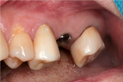
Tooth Site: #26
Diagnosis: Maxillary sinusitis
Clinical Summary
Maxillary sinusitis secondary to implant placement at #26 performed at an outside clinic.
Treatment Plan
1) Aspiration of purulent exudate at the #26 implant site using a syringe
2) Concurrent pharmacological management for sinusitis
Conclusion
A lateral window approach was performed using a round bur mounted on a surgical handpiece. Aspiration was performed through the lateral window, and Augmentin was prescribed. At the one-week follow-up, clinical improvement was noted. A CBCT scan taken two months later confirmed complete resolution of the sinusitis.
Case Presentation
Radiographic Findings & Diagnosis
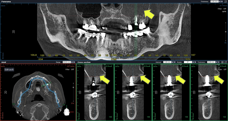
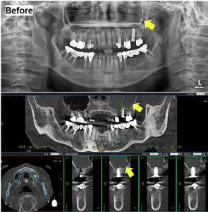
A panoramic radiograph revealed radiopacity within the left maxillary sinus. Clinical findings were consistent with implant-associated sinusitis at the #26 site.
Diagnosis: Implant-associated maxillary sinusitis


Treatment
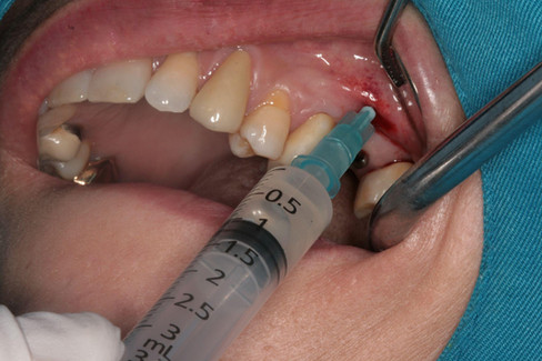
Following local anesthesia with one carpule of lidocaine, a minimally invasive incision was made, and a lateral window approach to the maxillary sinus was performed using a round bur.


Through the lateral window, purulent sinus exudate was aspirated using a 10cc syringe, resulting in a syringe filled with discharge.


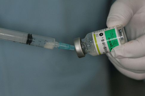
After aspiration, the maxillary sinus was irrigated with sterile saline, followed by direct injection of a solution containing 1g of Ceftizoxime sodium (Ceftezole sodium) mixed with 1cc of sterile distilled water into the sinus cavity.


Additionally, Augmentin was prescribed orally for 10 days to provide systemic antibiotic coverage.
Result
At the one-week follow-up, the lateral access site exhibited favorable clinical healing. The patient reported resolution of the previously noted malodor and overall symptomatic improvement. These findings suggest a favorable response to the initial treatment and a positive healing trajectory.
At the two-month follow-up, a panoramic radiograph and cone beam CT (CBCT) were obtained for comprehensive evaluation. Radiographic analysis showed that the prior radiopacity in the left maxillary sinus had resolved into radiolucency, suggesting resolution of the inflammation and restoration of sinus health.


Before & After


Clinical Video


