
- Neo Biotech
- Apr 2
- 2 min read

Updated: Apr 2

Prof. Richard Leesungbok (Korea)
Overview
This case presents immediate implant placement and loading in the mandibular anterior region at teeth #32 and #42 using a root-guided drilling approach. The treatment plan aimed to preserve socket architecture and optimize implant positioning by utilizing the remaining roots as a natural guide during pilot drilling.

Root Guide Surgery Procedure
Initial site preparation was performed directly over the remaining roots using a surgical bur. Pilot drilling with a 2.2 mm drill was then conducted, taking advantage of the root structure to guide the direction and depth of the osteotomy. This root-guided approach helped to maintain alignment with the original tooth trajectory, minimizing the risk of deviation. Drilling was continued sequentially up to 3.0 mm. Atraumatic root extraction was then performed to complete the site preparation.


Implant placement was performed in the prepared sites using Neobiotech IS-II implants (3.5 × 11.5 mm) at positions #32 and #42. An insertion torque of 40 Ncm was achieved, indicating excellent primary stability suitable for immediate loading. A temporary prosthesis was delivered on the same day to allow immediate loading, in accordance with the treatment plan.


Prosthetic Procedure
The surgical site and provisional restoration were stable on the first postoperative day, showing no signs of inflammation or complications.


Three month later, the provisional crown was removed, a transfer coping was connected, and a rubber impression was taken.


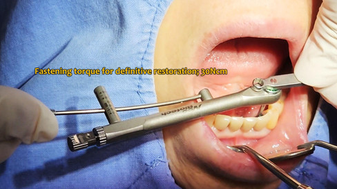
A working model was fabricated from the impression to proceed with the final prosthesis. Provisional restoration was removed before final prosthesis delivery. After verifying the fit, final PFM crowns were delivered and torqued to 30 Ncm to complete the prosthetic phase.


Follow-Up
At the 3-month follow-up, the peri-implant tissue and final restoration remained stable, with no clinical complications observed. By the 6-month follow-up, the patient continued to maintain a healthy intraoral condition, free of inflammation or prosthetic concerns. These findings indicate a favorable long-term prognosis in terms of both function and esthetics.
3-Month & 6-Month Follow-Up


Clinical Video


